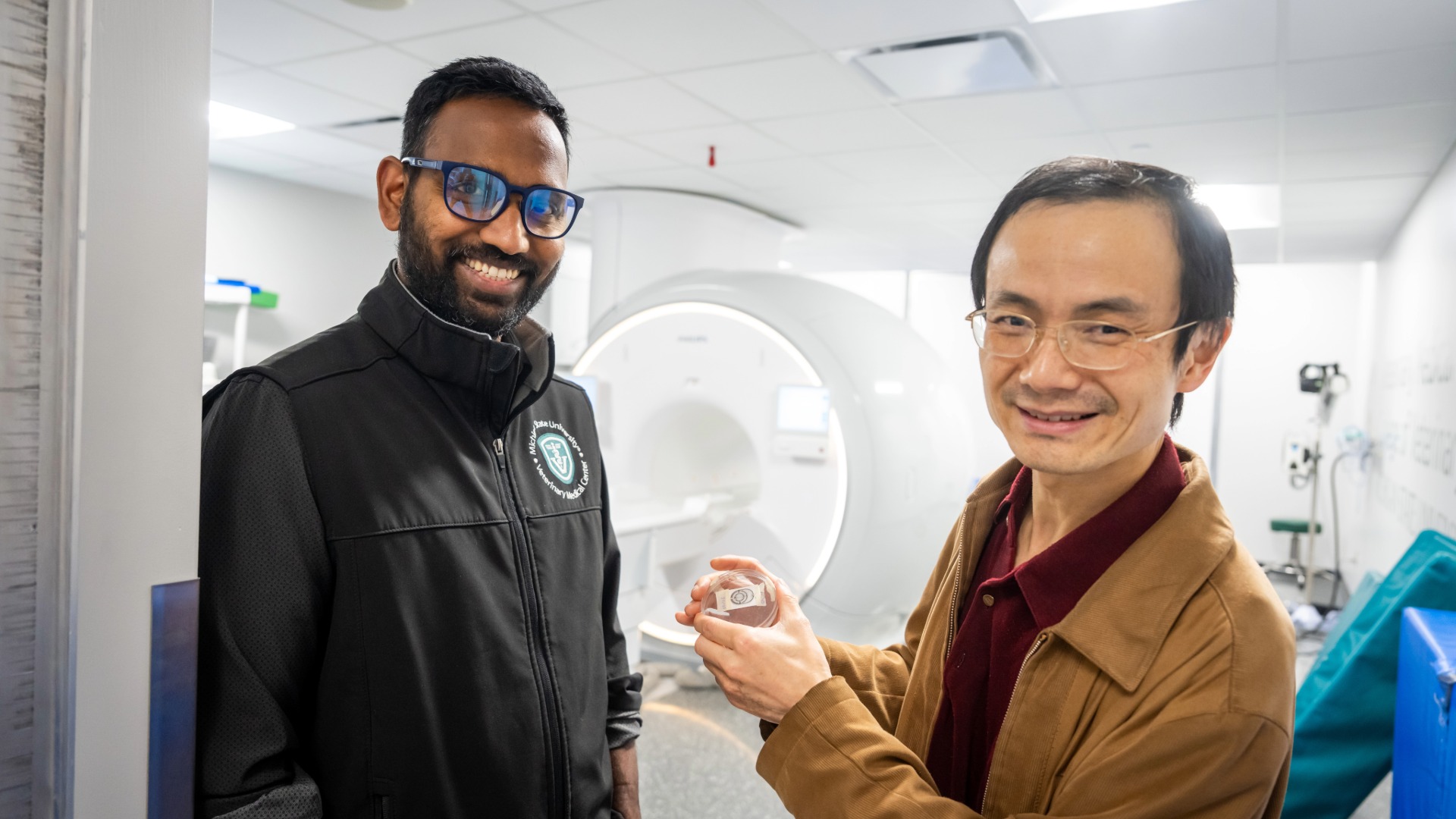
Chunqi Qian, Ph.D., associate professor in the Michigan State University Radiology Department with the College of Osteopathic Medicine (MSUCOM), is piloting research to develop a wireless, battery-free sensor that can detect early-stage changes in the brain and heart before symptoms become severe. Dr. Qian is leading efforts to use the device to measure abnormal activities in neurodegenerative diseases, including Parkinson’s and Alzheimer’s Diseases and cardiovascular diseases, such as arrythmia.
The sensor can reduce the need for expensive repetitive imaging like Magnetic Resonance Imaging (MRI), or it can be complementary by working inside an MRI to correlate signals without disturbing acquisition of the image. The sensor can also be used for remote and continuous monitoring of physiological parameters.
As described in WISDEM: a hybrid wireless integrated sensing detector for simultaneous EEG and MRI, published in Nature Methods, the sensor operates on radiofrequencies by harvesting energy from the ambient environment. What’s more, the sensor only needs a tiny amount of power to operate as an FM broadcaster, so it's a battery-free device which eliminates the need for battery replacement that could interrupt sensor recording.
When the sensor is placed inside an MRI machine, it uses the imaging scanner itself as a receiver. When the sensor is placed outside the scanner, it can also be conveniently detected by a low-cost FM receiver.
“In this scenario, the patient doesn't need to do repetitive scanning,” Dr. Qian said. “Instead, he or she only needs to go once, then go home and let the sensor operate itself for an extended period of time. Then if the sensor reports something unusual, that will trigger an alert for the patient to go back for more sophisticated imaging.”
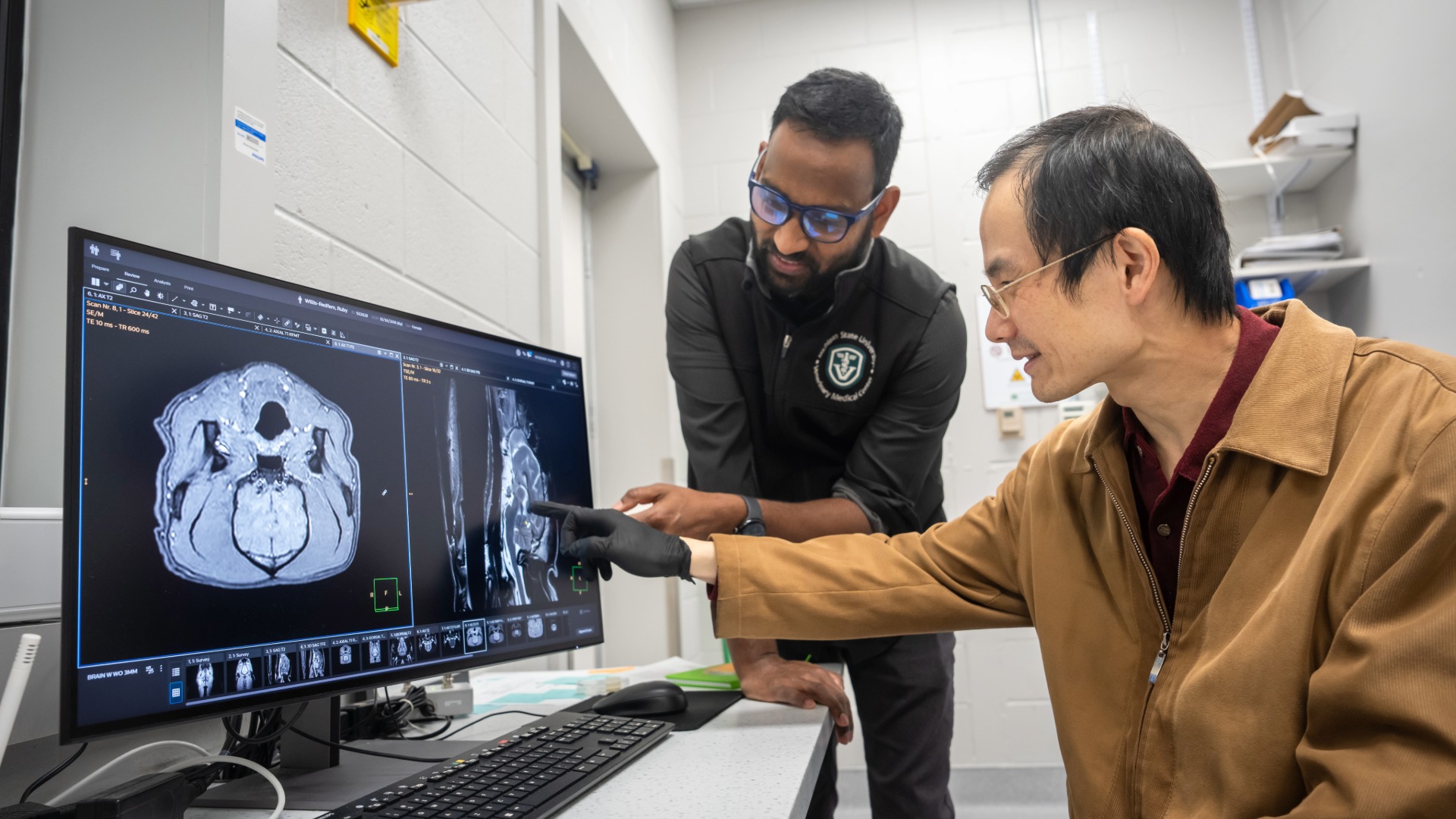
MRI visualizes vascular activities while specialized catheters or surface electrodes measure neuronal electrical firing. Patients can get MRI images or electrophysiology signals separately, but Dr. Qian wants to provide a convenient way to get them simultaneously inside an MRI scanner without complicated added-on apparatus.
Today, wired connections to electrodes are needed in conventional electrophysiology, but wired connections inside the MRI scanner can be problematic due to the electromagnetic interference. While there are techniques to synchronize the electrophysiology recording with MRI imaging, these techniques are complicated and have not been utilized clinically due to device complexity and safety concerns, such as the potential problem of overheating if wires are inside the MRI scanner.
The idea is to combine structural information from an MRI with the dynamic electrophysiology information recorded by the sensor in real time.
In addition to neurological disorders, Dr. Qian says the low-cost sensor could also be useful for cardiology applications such as diagnosing arrhythmia.
“Let's say a person goes to the hospital for an ECG (electrocardiogram) to check the heart,” he said. “The patient lies down and electrodes are put on the chest. But that's just a static case.
What we want to do is measure the patient's heart signal in real time -- when they’re working, sleeping, talking, when they’re doing many things. So, then you have to have a lightweight mobile sensor to record, upload and transmit all that information in real time.”
Animal testing and exploring commercialization pathways
Dr. Qian is collaborating with the MSU School of Veterinary Medicine to first test the wireless continuous sensor on companion animals. Owners of companion animals visiting MSU’s veterinary facilities must often make multiple trips to and from campus for repetitive ECG measurements, with each trip costing significant time and money.
“Existing sensors don’t send out data in real time,” Dr. Qian explained. “Instead, it's recorded locally on a sensor and then it has to be retrieved when the patient returns the sensor back to a veterinary hospital. So, we’re trying to overcome this limitation by configuring the sensor to broadcast all the time, enabling veterinarians to real-time monitor the pet’s health status for on-time intervention.”
Once the research device has been thoroughly tested in companion animals, Dr. Qian’s team can submit safety performance data to the Food and Drug Administration with the aim of expanding the sensor’s applications to human cardiology and other healthcare areas.
“Our long-term goal is to test them in humans and then to develop wearable, battery-free sensors for human applications, like extracting a complete waveform of the heart rate,” Dr. Qian said.
“That will be very valuable – not only for the heart application, because the heart signal is easier to identify -- but eventually we want to apply it all around the body, including the brain (EEG), eyes (EOG) and muscle (EMG). So, wherever an electric signal emits from the body, we can detect it and measure it in an MRI-compatible, battery-free way.”
An EEG (electroencephalogram) records the electrical activity of the brain, an EOG (electro-oculogram) is a test used to diagnose retinal diseases, and an EMG (electromyogram) is a medical procedure used to detect neuromuscular abnormalities.
This research was partially supported by an NIH BRAIN initiative grant. Right now, it is supported by a National Science Foundation (NSF) grant that is good through June 30, 2027. He is hoping to secure additional research funding and is also working with the MSU Research Foundation to explore commercialization pathways with Michigan Translational Research and Commercialization (MTRAC) business development grants or private investors. His research team is willing to distribute the sensors to corporate partners for testing and validation of their effectiveness in a community setting as a means of bringing the devices to the marketplace.
Dr. Qian is also exploring applications beyond the medical field, including agricultural and environmental uses for the sensor.
“For example, the sensor can track the position and physiology of an insect inside a bee colony, which could improve the efficiency of honey production,” Dr. Qian explained. “It could also be used for environmental monitoring, because the sensor can track environmental parameters, like temperature, humidity or chemical concentration, by wirelessly converting measurements into electric signals that can be detected and continuously broadcast by the sensor.”
By Lynn Waldsmith
Chunqi Qian, Ph.D., associate professor in the Michigan State University Radiology Department with the College of Osteopathic Medicine (MSUCOM), is piloting research to develop a wireless, battery-free sensor that can detect early-stage changes in the brain and heart before symptoms become severe. Dr. Qian is leading efforts to use the device to measure abnormal activities in neurodegenerative diseases, including Parkinson’s and Alzheimer’s Diseases and cardiovascular diseases, such as arrythmia.
The sensor can reduce the need for expensive repetitive imaging like Magnetic Resonance Imaging (MRI), or it can be complementary by working inside an MRI to correlate signals without disturbing acquisition of the image. The sensor can also be used for remote and continuous monitoring of physiological parameters.
As described in WISDEM: a hybrid wireless integrated sensing detector for simultaneous EEG and MRI, published in Nature Methods, the sensor operates on radiofrequencies by harvesting energy from the ambient environment. What’s more, the sensor only needs a tiny amount of power to operate as an FM broadcaster, so it's a battery-free device which eliminates the need for battery replacement that could interrupt sensor recording.
When the sensor is placed inside an MRI machine, it uses the imaging scanner itself as a receiver. When the sensor is placed outside the scanner, it can also be conveniently detected by a low-cost FM receiver.
“In this scenario, the patient doesn't need to do repetitive scanning,” Dr. Qian said. “Instead, he or she only needs to go once, then go home and let the sensor operate itself for an extended period of time. Then if the sensor reports something unusual, that will trigger an alert for the patient to go back for more sophisticated imaging.”
MRI visualizes vascular activities while specialized catheters or surface electrodes measure neuronal electrical firing. Patients can get MRI images or electrophysiology signals separately, but Dr. Qian wants to provide a convenient way to get them simultaneously inside an MRI scanner without complicated added-on apparatus.
Today, wired connections to electrodes are needed in conventional electrophysiology, but wired connections inside the MRI scanner can be problematic due to the electromagnetic interference. While there are techniques to synchronize the electrophysiology recording with MRI imaging, these techniques are complicated and have not been utilized clinically due to device complexity and safety concerns, such as the potential problem of overheating if wires are inside the MRI scanner.
The idea is to combine structural information from an MRI with the dynamic electrophysiology information recorded by the sensor in real time.
In addition to neurological disorders, Dr. Qian says the low-cost sensor could also be useful for cardiology applications such as diagnosing arrhythmia.
“Let's say a person goes to the hospital for an ECG (electrocardiogram) to check the heart,” he said. “The patient lies down and electrodes are put on the chest. But that's just a static case.
What we want to do is measure the patient's heart signal in real time -- when they’re working, sleeping, talking, when they’re doing many things. So, then you have to have a lightweight mobile sensor to record, upload and transmit all that information in real time.”
Animal testing and exploring commercialization pathways
Dr. Qian is collaborating with the MSU School of Veterinary Medicine to first test the wireless continuous sensor on companion animals. Owners of companion animals visiting MSU’s veterinary facilities must often make multiple trips to and from campus for repetitive ECG measurements, with each trip costing significant time and money.
“Existing sensors don’t send out data in real time,” Dr. Qian explained. “Instead, it's recorded locally on a sensor and then it has to be retrieved when the patient returns the sensor back to a veterinary hospital. So, we’re trying to overcome this limitation by configuring the sensor to broadcast all the time, enabling veterinarians to real-time monitor the pet’s health status for on-time intervention.”
Once the research device has been thoroughly tested in companion animals, Dr. Qian’s team can submit safety performance data to the Food and Drug Administration with the aim of expanding the sensor’s applications to human cardiology and other healthcare areas.
“Our long-term goal is to test them in humans and then to develop wearable, battery-free sensors for human applications, like extracting a complete waveform of the heart rate,” Dr. Qian said.
“That will be very valuable – not only for the heart application, because the heart signal is easier to identify -- but eventually we want to apply it all around the body, including the brain (EEG), eyes (EOG) and muscle (EMG). So, wherever an electric signal emits from the body, we can detect it and measure it in an MRI-compatible, battery-free way.”
An EEG (electroencephalogram) records the electrical activity of the brain, an EOG (electro-oculogram) is a test used to diagnose retinal diseases, and an EMG (electromyogram) is a medical procedure used to detect neuromuscular abnormalities.
This research was partially supported by an NIH BRAIN initiative grant. Right now, it is supported by a National Science Foundation (NSF) grant that is good through June 30, 2027. He is hoping to secure additional research funding and is also working with the MSU Research Foundation to explore commercialization pathways with Michigan Translational Research and Commercialization (MTRAC) business development grants or private investors. His research team is willing to distribute the sensors to corporate partners for testing and validation of their effectiveness in a community setting as a means of bringing the devices to the marketplace.
Dr. Qian is also exploring applications beyond the medical field, including agricultural and environmental uses for the sensor.
“For example, the sensor can track the position and physiology of an insect inside a bee colony, which could improve the efficiency of honey production,” Dr. Qian explained. “It could also be used for environmental monitoring, because the sensor can track environmental parameters, like temperature, humidity or chemical concentration, by wirelessly converting measurements into electric signals that can be detected and continuously broadcast by the sensor.”
By Lynn Waldsmith